

Imagine your arm suddenly goes weak while you are reading a book. You panic, but ten minutes later, it feels completely normal again. Did you just have a heart attack? No. But did you just ignore a critical warning from your brain? Many people think that if the symptoms go away, the danger is gone. This is a dangerous myth. A TIA is a transient episode of neurological dysfunction caused by focal brain, spinal cord, or retinal ischemia, without acute infarction. It is not a "mini-stroke" in the sense of being minor; it is a major red flag that demands immediate medical attention. The difference between a TIA and a full-blown stroke often comes down to time and tissue damage. While both conditions share identical warning signs, the outcome depends on whether blood flow was interrupted long enough to kill brain cells. Understanding this distinction is vital because the window for preventing a catastrophic stroke is incredibly narrow. For every person who experiences a TIA, the clock starts ticking immediately. The risk of having a major stroke within the next 90 days is as high as 20%, with the highest danger occurring in the first 48 hours.
Understanding the Core Difference: Tissue Damage vs. Temporary Blockage
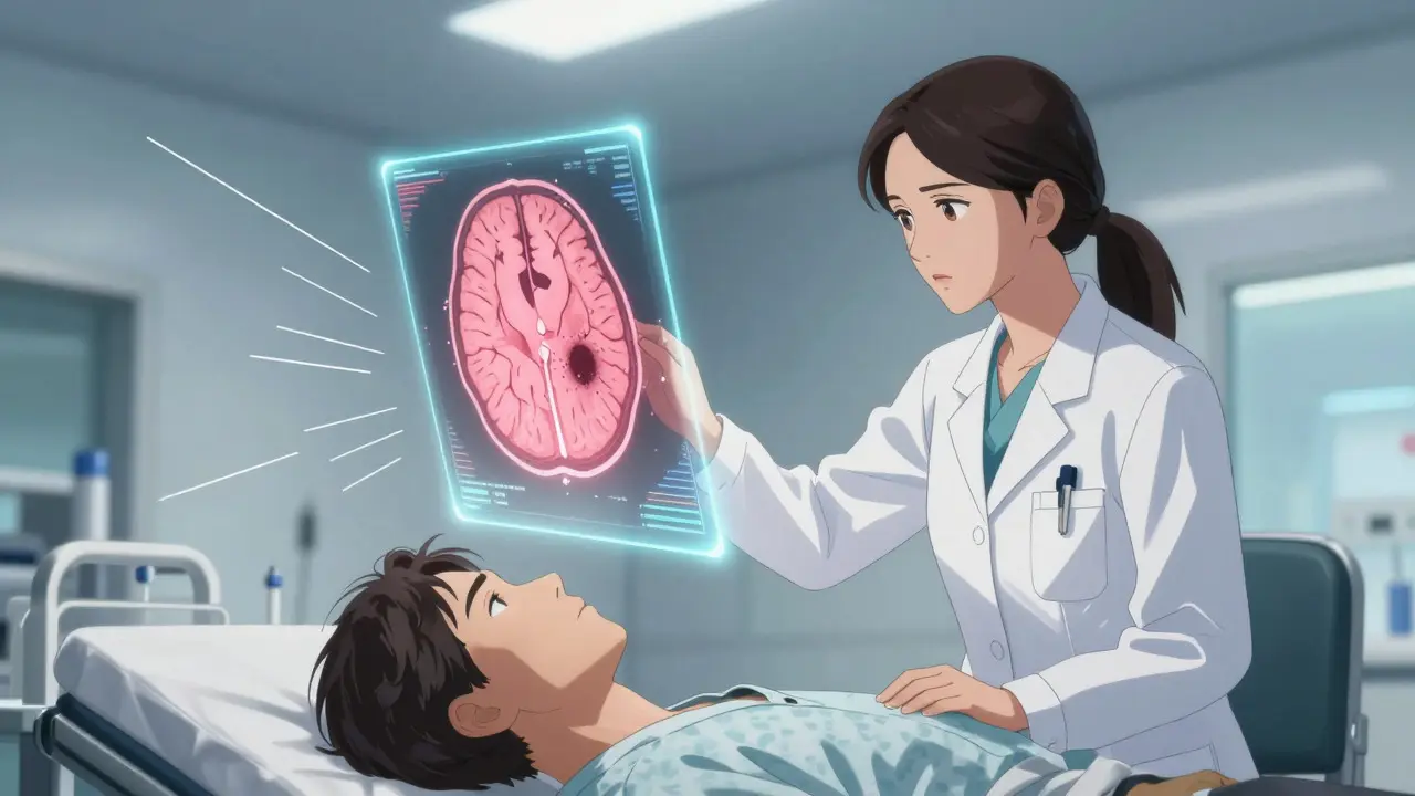
To grasp why urgency matters, we need to look at what is happening inside the brain. A Stroke is a condition where blood supply to part of the brain is cut off, causing permanent damage to brain tissue. When blood stops flowing, brain cells begin to die within minutes. This death creates an area of dead tissue called an infarction. On an MRI scan, this shows up clearly as a dark spot indicating permanent injury.
A TIA, on the other hand, involves a temporary blockage. Blood flow is interrupted, causing symptoms like weakness or speech difficulties, but the blockage clears before significant cell death occurs. Historically, doctors defined a TIA by how long the symptoms lasted (less than 24 hours). However, modern medicine has moved away from this time-based definition. Today, the gold standard for diagnosis is imaging. If a diffusion-weighted MRI shows no evidence of infarction, it is classified as a TIA. If it shows even a tiny spot of dead tissue, it is reclassified as a minor stroke, regardless of how quickly the symptoms resolved.
This shift in definition is crucial. Research indicates that approximately 35% of patients who present with suspected TIA symptoms actually show acute infarction on MRI. This means more than one in three "TIAs" are actually small strokes. Calling it a "mini-stroke" minimizes the reality that brain tissue may have been damaged. Dr. Steven Levine, a prominent neurologist, argues that a TIA is not just a warning but an actual stroke event that resolved spontaneously. This perspective changes how we treat these patients: they are not just at risk; they have already had a vascular event.
Recognizing the Warning Signs: BE FAST
The symptoms of a TIA and an ischemic stroke are virtually identical. This makes recognition tricky because you cannot tell them apart by looking at the patient alone. You need to rely on the pattern of onset and resolution. Both conditions strike suddenly, without gradual buildup. The most reliable way to identify these signs is through the BE FAST mnemonic, which expands on the traditional F.A.S.T. acronym to include balance and vision issues.
- B - Balance: Sudden loss of balance or coordination, such as stumbling or feeling dizzy without a clear reason.
- E - Eyes: Vision changes, including double vision, blurred vision, or loss of vision in one or both eyes.
- F - Face: Ask the person to smile. Does one side of the face droop? Is the smile uneven?
- A - Arms: Ask the person to raise both arms. Does one arm drift downward? Is there weakness or numbness in one arm or leg?
- S - Speech: Ask the person to repeat a simple sentence. Is their speech slurred, strange, or difficult to understand? Can they speak at all?
- T - Time: Note the time when symptoms first appeared. Call emergency services immediately.
It is important to note that these symptoms can affect only one side of the body. For example, right-sided weakness usually indicates a problem in the left hemisphere of the brain. The key differentiator in real-time observation is duration. TIA symptoms typically resolve within 10 to 60 minutes. In fact, 56% of TIAs resolve within one hour. If the symptoms persist beyond 24 hours, it is almost certainly a stroke. However, you should never wait 24 hours to see if it goes away. Waiting is the single biggest risk factor for a subsequent major stroke.
The Urgency Factor: Why Minutes Matter
Why is a TIA considered a medical emergency if the symptoms disappear? Because the underlying cause-usually a clot or plaque narrowing-is still there. The TIA is essentially a dry run. The American Heart Association designates TIA as a neurological emergency requiring evaluation within 24 hours. Ideally, assessment should happen within 60 minutes of symptom resolution. Every hour of delay increases the chance that the next blockage will be bigger and last longer.
Data from the EXPRESS study published in *The Lancet* showed that immediate treatment within 24 hours reduced the 90-day stroke risk from 10.3% to 2.1%. That is a massive drop. Conversely, delayed care leads to higher conversion rates. Hospitals that fail to act quickly see significantly more TIA patients go on to suffer full strokes. The Centers for Medicare & Medicaid Services now penalize hospitals with high TIA-to-stroke conversion rates, driving stricter compliance with rapid evaluation protocols.
Public misconception plays a huge role in this delay. About 31% of TIA patients wait more than 24 hours to seek care, often because they feel fine again. This "wait and see" approach is statistically risky. The risk of stroke after a TIA is not evenly distributed over 90 days; it is front-loaded. The first 48 hours carry the highest danger. If you miss this window, you are gambling with a system that has already shown it can fail.
Risk Stratification: The ABCD2 Score
Once a patient arrives at the emergency department, doctors need a quick way to determine how urgent the situation is. They use the ABCD2 scoring system, a validated tool that quantifies the risk of stroke in the next two days. It assigns points based on five factors:
- A - Age: 1 point if age is 60 or older.
- B - Blood Pressure: 1 point if systolic BP is 140 mmHg or higher, or diastolic is 90 mmHg or higher.
- C - Clinical Features: 2 points for unilateral weakness; 1 point for speech impairment without weakness.
- D - Duration: 2 points if symptoms lasted 60 minutes or more; 1 point if 10-59 minutes.
- D - Diabetes: 1 point if the patient has diabetes.
The total score ranges from 0 to 7. A score of 4 or higher indicates a high risk of stroke within 48 hours (approximately 8.1%). Patients with high scores are typically admitted to the hospital for closer monitoring and faster intervention. Those with lower scores might be discharged with strict follow-up instructions, but they still require urgent outpatient evaluation. This scoring system helps triage resources, ensuring that those most likely to have a second event get the fastest care.
Diagnostic Pathway: From CT to MRI
The diagnostic process follows a specific sequence designed to rule out life-threatening complications first. The first step is always a non-contrast CT scan of the head. This does not show early ischemic strokes well, but it is excellent at ruling out hemorrhagic strokes (bleeding in the brain). Since bleeding requires different treatment (often surgery or reversal of blood thinners), this step is critical before giving any medication.
After the CT, the patient needs an MRI with diffusion-weighted imaging (DWI). DWI is highly sensitive to acute infarction, detecting cell death within minutes. It is the definitive test to distinguish TIA from minor stroke. If the DWI is negative, the diagnosis remains TIA. If positive, it is a minor stroke. Beyond imaging, doctors look for the source of the blockage. Common causes include carotid artery stenosis (narrowing) or atrial fibrillation (an irregular heartbeat that throws clots to the brain). Carotid ultrasound and echocardiograms are standard tests to find these sources.
| Feature | TIA (Transient Ischemic Attack) | Ischemic Stroke |
|---|---|---|
| Brain Tissue Damage | No infarction on MRI | Permanent infarction visible on MRI |
| Symptom Duration | Typically resolves in <1 hour (up to 24h historically) | Persists >24 hours or causes permanent deficit |
| Primary Risk | High risk of future major stroke (20% in 90 days) | Immediate disability, death, or secondary complications |
| Immediate Treatment Focus | Rapid risk stratification, antiplatelet therapy | Reperfusion (clot-busting drugs/thrombectomy) |
| Prognosis | Good if treated promptly; prevents future stroke | Depends on size/location of infarction; often leaves deficits |
Treatment and Prevention Strategies
Treating a TIA is about preventing the next event. Unlike a large stroke, where the goal is to save dying brain tissue, TIA management focuses on secondary prevention. The cornerstone of treatment is antiplatelet therapy. Aspirin 325 mg is often started within 24 hours. For high-risk patients, dual antiplatelet therapy (aspirin plus clopidogrel) is used for 21 to 30 days to reduce the risk of another clot forming. This combination has been shown to reduce subsequent stroke risk significantly compared to aspirin alone.
Beyond blood thinners, controlling cardiovascular risk factors is essential. High-intensity statins, such as atorvastatin 80 mg daily, are prescribed to stabilize plaques in the arteries. Blood pressure must be managed strictly, aiming for levels below 140/90 mmHg. If the TIA was caused by atrial fibrillation, anticoagulants like warfarin or direct oral anticoagulants (DOACs) are used instead of antiplatelets, as they are more effective at preventing cardioembolic clots.
Lifestyle changes also play a role, though they are slower acting. Quitting smoking, managing diabetes, and adopting a heart-healthy diet contribute to long-term stability. However, these changes do not replace the need for immediate pharmacological intervention. The goal is to keep the arterial system clean and the heart rhythm regular so that no further emboli reach the brain.
Common Misconceptions and Pitfalls
The biggest enemy in TIA management is complacency. People often think, "It went away, so I'm fine." This mindset delays care and increases mortality. Another common pitfall is attributing symptoms to benign causes like vertigo, anxiety, or low blood sugar. While these can mimic TIA symptoms, the sudden onset and focal nature (one side of the body) of TIA symptoms make them distinct. Dizziness alone is less specific, but dizziness accompanied by facial drooping or arm weakness is a red flag.
There is also confusion about the term "mini-stroke." While widely used, it implies the event is small and manageable. In reality, a TIA is a major neurological event with a high probability of leading to a disabling stroke. Patients need to understand that the TIA itself is the crisis, not just a precursor. Finally, some patients skip follow-up appointments because they feel well. Adherence to medication and regular check-ups are critical. Missing doses of antiplatelets or statins can allow plaque to destabilize, increasing the risk of a breakthrough stroke.
Next Steps for Suspected TIA
If you or someone else experiences sudden neurological symptoms, do not wait. Even if the symptoms fade, call emergency services. Time is brain. In the emergency room, expect a rapid workup including a CT scan, blood tests, and possibly an MRI. Be prepared to answer questions about the exact timing of symptoms, medical history, and current medications. After discharge, ensure you have a plan for follow-up with a neurologist. Keep a log of any recurring symptoms, no matter how brief. With proper evaluation and treatment, the vast majority of TIA patients can prevent a major stroke and return to their normal lives. The key is speed and vigilance.
What is the difference between a TIA and a mini-stroke?
Medically, there is no difference; "mini-stroke" is a lay term for TIA. However, experts discourage the term because it minimizes the severity. A TIA is a serious warning sign with a high risk of leading to a major stroke, not a minor health issue.
How long do TIA symptoms usually last?
Most TIA symptoms resolve within 10 to 60 minutes. About 56% of cases resolve within one hour. Historically, the limit was set at 24 hours, but modern diagnosis relies on imaging rather than time.
Do I need to go to the hospital if my symptoms go away?
Yes. A TIA is a medical emergency. You need urgent evaluation to start preventive treatment and assess your risk of a future stroke. Delaying care increases the likelihood of a major stroke within the next 48 hours.
What tests are done to diagnose a TIA?
Doctors typically perform a CT scan to rule out bleeding, followed by an MRI with diffusion-weighted imaging to check for tissue damage. They may also use carotid ultrasounds and echocardiograms to find the source of the blockage.
Can a TIA happen more than once?
Yes, recurrent TIAs are possible, especially if the underlying cause is not treated. Each TIA increases the risk of a major stroke, making consistent medication adherence and lifestyle changes critical for prevention.